Custom YOLO Object Detection Model for COVID-19 Diagnosis
DOI:
https://doi.org/10.51173/jt.v5i3.1174Keywords:
Yolo, Convolutional Neural Network, Coronavirus, Object DetectionAbstract
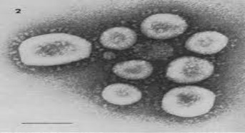
The emergence and spread of the new coronavirus (COVID-19) poses a new public health threat to the entire world (SARS-CoV-2). This new virus is highly contagious and pathogenetically different from other mainstream respiratory viruses. Clinical staff can benefit from Computer Aided Diagnostics (CAD) systems that combine deep learning algorithms and image processing technologies as diagnostic tools for COVID-19. These tools also help to better understand the course of the disease. In most cases, medical staff and healthcare facilities would be more equipped to promptly diagnose COVID-19 for patients with improved flexibility. To examine the training performance of the contemporary YOLOv4 model, this work presents the development of a computer-assisted automatic detection system that focuses specifically on identifying viral cells in blood samples from patients using electron microscopy images to detect the infected blood cell. The mean average precision of the proposed custom model is 86.5%mAP, making it suitable for the upcoming COVID-19 monitoring systems.
Downloads
References
Guo, H., et al., The impact of the COVID-19 epidemic on the utilization of emergency dental services. Journal of dental sciences, 2020. 15(4): p. 564-567.
Lundervold, A.S. and A. Lundervold, An overview of deep learning in medical imaging focusing on MRI. Zeitschrift für Medizinische Physik, 2019. 29(2): p. 102-127.
Chen, Y., et al., New-onset autoimmune phenomena post-COVID-19 vaccination. Immunology, 2022. 165(4): p. 386-401.
Redmon, J. and A. Farhadi. YOLO9000: better, faster, stronger. in Proceedings of the IEEE conference on computer vision and pattern recognition. 2017.
Martin-Cardona, A., et al., SARS-CoV-2 identified by transmission electron microscopy in lymphoproliferative and ischaemic intestinal lesions of COVID-19 patients with acute abdominal pain: two case reports. BMC gastroenterology, 2021. 21(1): p. 1-10.
Redmon, J., et al. You only look once: Unified, real-time object detection. in Proceedings of the IEEE conference on computer vision and pattern recognition. 2016.
Redmon, J. and A. Farhadi, Yolov3: An incremental improvement. arXiv preprint arXiv:1804.02767, 2018.
Girshick, R., et al. Rich feature hierarchies for accurate object detection and semantic segmentation. in Proceedings of the IEEE conference on computer vision and pattern recognition. 2014.
LeCun, Y., Y. Bengio, and G. Hinton, Deep learning. nature, 2015. 521(7553): p. 436-444.
Liu, W., et al. Ssd: Single shot multibox detector. in European conference on computer vision. 2016. Springer.
Anwar, S.M., et al., Medical image analysis using convolutional neural networks: a review. Journal of medical systems, 2018. 42(11): p. 1-13.
Ker, J., et al., Deep learning applications in medical image analysis. Ieee Access, 2017. 6: p. 9375-9389.
Singh, S.P., et al., 3D deep learning on medical images: a review. Sensors, 2020. 20(18): p. 5097.
Ker, J., et al., Image thresholding improves 3-dimensional convolutional neural network diagnosis of different acute brain hemorrhages on computed tomography scans. Sensors, 2019. 19(9): p. 2167.
Singh, S.P., et al., Shallow 3D CNN for detecting acute brain hemorrhage from medical imaging sensors. IEEE Sensors Journal, 2020. 21(13): p. 14290-14299.
Yadav, S.S. and S.M. Jadhav, Deep convolutional neural network based medical image classification for disease diagnosis. Journal of Big Data, 2019. 6(1): p. 1-18.
Milica, M. and M.C. Badža, BarjaktaroviC. Classification of Brain Tumors from MRI Images Using a Convolutional Neural Network. MDPI. 2020, MultiDisciplinary Digital Publishing institute.
Ker, J., et al., Automated brain histology classification using machine learning. Journal of Clinical Neuroscience, 2019. 66: p. 239-245.
Zhou, T., S. Ruan, and S. Canu, A review: Deep learning for medical image segmentation using multi-modality fusion. Array, 2019. 3: p. 100004.
Ramesh, K., et al., A review of medical image segmentation algorithms. EAI Endorsed Transactions on Pervasive Health and Technology, 2021. 7(27): p. e6-e6.
Nair, R., et al., Detection of COVID-19 cases through X-ray images using hybrid deep neural network. World Journal of Engineering, 2022. 19(1): p. 33-39.
Medhi, K., M. Jamil, and M.I. Hussain, Automatic detection of COVID-19 infection from chest X-ray using deep learning. medrxiv, 2020: p. 2020.05. 10.20097063.
Sethy, P.K. and S.K. Behera, Detection of coronavirus disease (covid-19) based on deep features. 2020.
Apostolopoulos, I.D. and T.A. Mpesiana, Covid-19: automatic detection from x-ray images utilizing transfer learning with convolutional neural networks. Physical and engineering sciences in medicine, 2020. 43: p. 635-640.
Xu, X., et al., A deep learning system to screen novel coronavirus disease 2019 pneumonia. Engineering, 2020. 6(10): p. 1122-1129.
Hussain, E., et al., CoroDet: A deep learning based classification for COVID-19 detection using chest X-ray images. Chaos, Solitons & Fractals, 2021. 142: p. 110495.
Ozturk, T., et al., Automated detection of COVID-19 cases using deep neural networks with X-ray images. Computers in biology and medicine, 2020. 121: p. 103792.
Bullock, H.A., C.S. Goldsmith, and S.E. Miller, Best practices for correctly identifying coronavirus by transmission electron microscopy. Kidney International, 2021. 99(4): p. 824-827.
Koylu, C., C. Zhao, and W. Shao, Deep neural networks and kernel density estimation for detecting human activity patterns from geo-tagged images: A case study of birdwatching on flickr. ISPRS international journal of geo-information, 2019. 8(1): p. 45.
Wang, C.-Y., et al. CSPNet: A new backbone that can enhance learning capability of CNN. in Proceedings of the IEEE/CVF conference on computer vision and pattern recognition workshops. 2020.
Liu, S., et al. Path aggregation network for instance segmentation. in Proceedings of the IEEE conference on computer vision and pattern recognition. 2018.
Downloads
Published
How to Cite
Issue
Section
License
Copyright (c) 2023 Noor Najah Ali, Aseel Hameed, Ali Al_Naji

This work is licensed under a Creative Commons Attribution 4.0 International License.
