
Calcium Phosphate Coatings with Controlled Micro/Nano-Structures for Endothelial Cells Viability
DOI:
https://doi.org/10.51173/jt.v6i2.2545Keywords:
Hydroxyapatite, Micro/Nano-Structure, Scaffolds, Coating, Endothelial CellAbstract
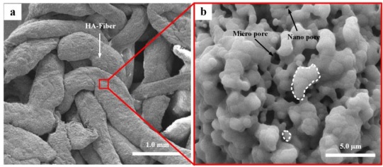
Hydroxyapatite (HA) scaffolds produced by the accumulation of HA fibers were separately treated hydrothermally in three calcium phosphate solutions to form coatings of different micro/nano-structures. Different micro/nano-structure and morphologies have been regulated on the surface of treated HA scaffolds. Plate-like compromise flower-like morphology was obtained with solution 1 (Ca-sufficient) i.e., ratio: Ca/Ca=1%; Ca/P=1.67. Full coatings (flower-like) morphology treated after Cu-doped coating solution 2 (Cu/(Cu+Ca) = 5%; ratio: (Cu+Ca)/P = 1.67). Furthermore, partial coatings (flower-like) morphology fabricated with solution 3 (Ca-deficient and Cu-replacement), i.e., ratio: Ca/Ca=0.95%; Ca/P=1.58. The results showed the effect of hydrothermal coatings on HA scaffolds. Cultured human endothelial cells spread and proliferated better on the treated HA scaffolds than on the uncoated scaffolds, suggesting a potential effect of calcium phosphate surface morphology on endothelial cell response. Thus, it can provide an appropriate micro/nano-structure approach supporting angiogenesis capacity, which is a necessity to accelerate the time of bone healing and regeneration.
Downloads
References
N. Eliaz and N. Metoki, "Calcium Phosphate Bioceramics: A Review of Their History, Structure, Properties, Coating Technologies and Biomedical Applications," Materials, vol. 10, no. 4, 2017, https://doi.org/10.3390/ma10040334.
T. M. Sridhar, U. K. Mudali, and M. Subbaiyan, "Preparation and characterisation of electrophoretically deposited hydroxyapatite coatings on type 316L stainless steel," Corrosion Science, vol. 45, no. 2, pp. 237-252, 2003, https://doi.org/10.1016/S0010-938X(02)00091-4.
S. S. A. Abidi and Q. Murtaza, "Synthesis and Characterization of Nano-hydroxyapatite Powder Using Wet Chemical Precipitation Reaction," Journal of Materials Science & Technology, vol. 30, no. 4, pp. 307-310, 2014, https://doi.org/10.1016/j.jmst.2013.10.011.
K. Batebi, B. Abbasi Khazaei, and A. Afshar, "Characterization of sol-gel derived silver/fluor-hydroxyapatite composite coatings on titanium substrate," Surface and Coatings Technology, vol. 352, pp. 522-528, 2018, https://doi.org/10.1016/j.surfcoat.2018.08.021 .
M. Othmani, H. Bachoua, Y. Ghandour, A. Aissa, and M. Debbabi, "Synthesis, characterization and catalytic properties of copper-substituted hydroxyapatite nanocrystals," Materials Research Bulletin, vol. 97, pp. 560-566, 2018, https://doi.org/10.1016/j.materresbull.2017.09.056.
D. Xiao, T. Guo, F. Yang, G. Feng, F. Shi, J. Li, et al., "In situ formation of nanostructured calcium phosphate coatings on porous hydroxyapatite scaffolds using a hydrothermal method and the effect on mesenchymal stem cell behavior," Ceramics International, vol. 43, no. 1, pp. 1588-1596, 2017, https://doi.org/10.1016/j.ceramint.2016.10.023.
M. M. Taheri, M. R. Abdul Kadir, T. Shokuhfar, A. Hamlekhan, M. Assadian, M. R. Shirdar, et al., "Surfactant-assisted hydrothermal synthesis of Fluoridated Hydroxyapatite nanorods," Ceramics International, vol. 41, no. 8, pp. 9867-9872, 2015, https://doi.org/10.1016/j.ceramint.2015.04.061.
X. Ye, C. Zhou, Z. Xiao, Y. Fan, X. Zhu, Y. Sun, et al., "Fabrication and characterization of porous 3D whisker-covered calcium phosphate scaffolds," Materials Letters, vol. 128, pp. 179-182, 2014, https://doi.org/10.1016/j.matlet.2014.04.142 .
J. Qin, Z. Zhong, and J. Ma, "Biomimetic synthesis of hybrid hydroxyapatite nanoparticles using nanogel template for controlled release of bovine serum albumin," Mater Sci Eng C Mater Biol Appl, vol. 62, pp. 377-83, May 2016, https://doi.org/10.1016/j.msec.2016.01.088.
A. Elrayah, W. Zhi, S. Feng, S. Al-Ezzi, H. Lei, and J. Weng, "Preparation of Micro/Nano-Structure Copper-Substituted Hydroxyapatite Scaffolds with Improved Angiogenesis Capacity for Bone Regeneration," Materials (Basel), vol. 11, no.9, Aug 23 2018, https://doi.org/10.3390/ma11091516.
S. Guang, F. Ke, and Y. Shen, "Controlled Preparation and Formation Mechanism of Hydroxyapatite Nanoparticles under Different Hydrothermal Conditions," Journal of Materials Science & Technology, vol. 31, no.8, pp. 852-856, 2015, https://doi.org/10.1016/j.jmst.2014.12.013.
S. M. Chim, J. Tickner, S. T. Chow, V. Kuek, B. Guo, G. Zhang, et al., "Angiogenic factors in bone local environment," Cytokine Growth Factor Rev, vol. 24, no. 3, pp. 297-310, Jun 2013, https://doi.org/10.1016/j.cytogfr.2013.03.008.
M. S. Laranjeira, M. H. Fernandes, and F. J. Monteiro, "Response of monocultured and co-cultured human microvascular endothelial cells and mesenchymal stem cells to macroporous granules of nanostructured-hydroxyapatite agglomerates," J Biomed Nanotechnol, vol. 9, no. 9, pp. 1594-606, Sep 2013, https://doi.org/10.1166/jbn.2013.1664.
S. Pezzatini, R. Solito, L. Morbidelli, S. Lamponi, E. Boanini, A. Bigi, et al., "The effect of hydroxyapatite nanocrystals on microvascular endothelial cell viability and functions," J Biomed Mater Res A, vol. 76, no.3, pp. 656-63, Mar 1 2006, https://doi.org/10.1002/jbm.a.30524.
G.-S. Lee, J.-H. Park, U. S. Shin, and H.-W. Kim, "Direct deposited porous scaffolds of calcium phosphate cement with alginate for drug delivery and bone tissue engineering," Acta biomaterialia, vol. 7, no.8, pp. 3178-3186, 2011, https://doi.org/10.1016/j.actbio.2011.04.008.
Z. S. Stojanovic, N. Ignjatovic, V. Wu, V. Zunic, L. Veselinovic, S. Skapin, et al., "Hydrothermally processed 1D hydroxyapatite: Mechanism of formation and biocompatibility studies," Mater Sci Eng C Mater Biol Appl, vol. 68, pp. 746-57, Nov 01 2016, https://doi.org/10.1016/j.msec.2016.06.047 .
X. X. Wang, L. Yang, H. Liu, Q. Chen, D. Q. Xiao, and J. G. Zhu, "Optical Properties of ZnS:Co+Cr Nanocrystals Synthesized by a Low Temperature Hydrothermal Process," Journal of Inorganic Materials, vol. 29, no.10, pp. 1049-1054, 2014.
A. Magnaudeix, J. Usseglio, M. Lasgorceix, F. Lalloue, C. Damia, J. Brie, et al., "Quantitative analysis of vascular colonisation and angio-conduction in porous silicon-substituted hydroxyapatite with various pore shapes in a chick chorioallantoic membrane (CAM) model," Acta Biomaterialia, vol. 38, pp. 179-189, 2016, https://doi.org/10.1016/j.actbio.2016.04.039.
X. Li, C. A. van Blitterswijk, Q. Feng, F. Cui, and F. Watari, "The effect of calcium phosphate microstructure on bone-related cells in vitro," Biomaterials, vol. 29, no.23, pp. 3306-3316, 2008, https://doi.org/10.1016/j.biomaterials.2008.04.039.
F. E Imrie and J. M. S. Skakle, "Preparation of Copper-Doped Hydroxyapatite with Varying x in the Composition Ca10(PO4)6CuxOyHz," Bioceramics Development and Applications, vol. 3, 2013, DOI: 10.4172/2090-5025.S1-005.
M. A. Saghiri, A. Asatourian, J. Orangi, C. M. Sorenson, and N. Sheibani, "Functional role of inorganic trace elements in angiogenesis-Part II: Cr, Si, Zn, Cu, and S," Crit Rev Oncol Hematol, vol. 96, no.1, pp. 143-55, Oct 2015, https://doi.org/10.1016/j.critrevonc.2015.05.011.
Downloads
Published
How to Cite
Issue
Section
License
Copyright (c) 2024 Adil Elrayah, Ke Duan, Xiong Lu, Xiaob Lu, Jie Weng

This work is licensed under a Creative Commons Attribution 4.0 International License.