
A Novel Approach to Electrophoretic Deposition of Bioactive Glass on Ti-6Al-7Nb Using TEA-Stabilized Suspension
DOI:
https://doi.org/10.51173/jt.v7i4.2713Keywords:
Bioactive Glass, Triethanolamine, Electrophoretic Deposition Ti-6Al-7Nb Alloy, Zeta Potential, Corrosion Resistance, Adhesion StrengthAbstract
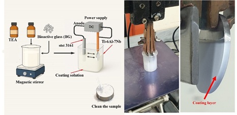
This study investigated the effect of varying bioactive glass (BG) concentrations (8–14 g/L) on the structural, electrochemical, and adhesive properties of coatings deposited on Ti-6Al-7Nb substrates via electrophoretic deposition (EPD). The SEM and EDS analyses demonstrated that rising BG concentration from 8 g/L to 14 g/L enhanced the coating uniformity, reduced the porosity and enhanced the particle packing. The coating thickness increased from 41.1 µm to 57.3 µm. Zeta potential measurements showed increased suspension stability with higher BG content, reaching 18.47 mV at 14 g/L. Corrosion resistance is enhanced with BG content, with the 14 g/L coating reaching the lowest corrosion current density (5.19 × 10⁻⁸ A) and rate (4.509 × 10⁻⁴ mmpy). Adhesion tests rated the 14 g/L BG coating as 1B due to better interlocking.
Downloads
References
O. Demontiero, C. Vidal, and G. Duque, “Aging and bone loss: New insights for the clinician,” Ther. Adv. Musculoskelet. Dis., vol. 4, no. 2, pp. 61–76, 2012, doi: 10.1177/1759720X11430858.
R. Drevet and H. Benhayoune, Biomaterials Design for Human Body Repair. Basel, Switzerland: MDPI, 2024.
G. Li et al., “An overview of osteoporosis and frailty in the elderly,” BMC Musculoskelet. Disord., vol. 18, Art. no. 46, 2017, doi: 10.1186/s12891-017-1411-3.
K. Moghadasi et al., “A review on biomedical implant materials and the effect of friction stir based techniques on their mechanical and tribological properties,” J. Mater. Res. Technol., vol. 17, pp. 1054–1121, 2022, doi: 10.1016/j.jmrt.2022.01.061.
G. Renganathan, N. Tanneru, and S. L. Madurai, “Orthopedical and biomedical applications of titanium and zirconium metals,” in Fundamental Biomaterials: Metals. Oxford, UK: Elsevier, 2018, pp. 211–241.
S. Mutha, “Evolution and principles of metals and alloys used in orthopedic implantology,” in Handbook of Orthopaedic Trauma Implantology. Cham, Switzerland: Springer, 2022, pp. 1–19.
J. Zhou et al., “Evolution from bioinert to bioresorbable: In vivo comparative study of additively manufactured metal bone scaffolds,” Adv. Sci., vol. 10, no. 26, Art. no. 2302702, 2023, doi: 10.1002/advs.202302702.
R. M. Pilliar, “Metallic biomaterials,” in Biomedical Materials. Boca Raton, FL, USA: CRC Press, 2009, pp. 41–81.
K. Prasad et al., “Metallic biomaterials: Current challenges and opportunities,” Materials, vol. 10, no. 8, Art. no. 884, 2017, doi: 10.3390/ma10080884.
D. Zindani, K. Kumar, and J. P. Davim, “Metallic biomaterials—A review,” in Mechanical Behaviour of Biomaterials. Sawston, UK: Woodhead Publishing, 2019, pp. 83–99.
J. Huang, X. Li, and Z. Guo, “Biomechanical and biochemical compatibility in innovative biomaterials,” in Biocompatibility and Performance of Medical Devices. Oxford, UK: Elsevier, 2020, pp. 23–46.
M. A. Shayeb et al., “Bioactive surface modifications on dental implants: A systematic review and meta-analysis,” Clin. Oral Investig., vol. 28, no. 11, Art. no. 592, 2024, doi: 10.1007/s00784-024-05892-1.
G. Zhu, G. Wang, and J. J. Li, “Advances in implant surface modifications to improve osseointegration,” Mater. Adv., vol. 2, no. 21, pp. 6901–6927, 2021, doi: 10.1039/D1MA00507A.
Y. Liu et al., “Role of implant surface modification in osseointegration: A systematic review,” J. Biomed. Mater. Res. A, vol. 108, no. 3, pp. 470–484, 2020, doi: 10.1002/jbm.a.36844.
D. S. Brauer, “Bioactive glasses—Structure and properties,” Angew. Chem. Int. Ed., vol. 54, no. 14, pp. 4160–4181, 2015, doi: 10.1002/anie.201405310.
A. Hoppe, N. S. Güldal, and A. R. Boccaccini, “A review of the biological response to ionic dissolution products from bioactive glasses and glass-ceramics,” Biomaterials, vol. 32, no. 11, pp. 2757–2774, 2011, doi: 10.1016/j.biomaterials.2011.01.004.
P. A. Hassan, B. Balakrishnan, and A. K. Tyagi, Engineered Biomaterials: Progress and Prospects. Singapore: Springer, 2023.
N. O. Joy-Anne et al., “Bioactive glass coatings on metallic implants for biomedical applications,” Bioact. Mater., vol. 4, pp. 261–270, 2019, doi: 10.1016/j.bioactmat.2019.06.003.
A. R. Boccaccini et al., “Polymer/bioactive glass nanocomposites for biomedical applications: A review,” Compos. Sci. Technol., vol. 70, no. 13, pp. 1764–1776, 2010, doi: 10.1016/j.compscitech.2010.06.002.
S. Albayrak and C. Gul, “Ceramic coatings for biomedical applications,” in Fiber and Ceramic Filler-Based Polymer Composites for Biomedical Engineering. Cham, Switzerland: Springer, 2024, pp. 233–256.
N. Nikam et al., “Advancements in surface coatings for enhancing longevity in hip implants: A review,” Prosthesis, vol. 7, no. 1, Art. no. 21, 2025, doi: 10.3390/prosthesis7010021.
H. Meskher et al., “Limitations, challenges and prospective solutions for bioactive glass-based nanocomposites for dental applications,” J. Dent., Art. no. 105331, 2024, doi: 10.1016/j.jdent.2024.105331.
A. Sola et al., “Bioactive glass coatings: A review,” Surf. Eng., vol. 27, no. 8, pp. 560–572, 2011, doi: 10.1179/1743294411Y.0000000024.
G. Brunello, H. Elsayed, and L. Biasetto, “Bioactive glass and silicate-based ceramic coatings on metallic implants,” Materials, vol. 12, no. 18, Art. no. 2929, 2019, doi: 10.3390/ma12182929.
R. Sergi, D. Bellucci, and V. Cannillo, “A comprehensive review of bioactive glass coatings,” Coatings, vol. 10, no. 8, Art. no. 757, 2020, doi: 10.3390/coatings10080757.
N. Eliaz and N. Metoki, “Calcium phosphate bioceramics,” Materials, vol. 10, no. 4, Art. no. 334, 2017, doi: 10.3390/ma10040334.
M. Maximov et al., “Bioactive glass—Preparation and coating methods,” Coatings, vol. 11, no. 11, Art. no. 1386, 2021, doi: 10.3390/coatings11111386.
R. Drevet, J. Fauré, and H. Benhayoune, “Electrophoretic deposition of bioactive glass coatings for bone implant applications,” Coatings, vol. 14, no. 9, Art. no. 1084, 2024, doi: 10.3390/coatings14091084.
N. I. Alyousef, A. T. Shahd, and A. S. Khana, “Bioactive glass coated dental implants,” in Dental Implants: Materials, Coatings, Surface Modifications and Interfaces with Oral Tissues. Cambridge, UK: Woodhead Publishing, 2020, pp. 93–112.
R. Drevet, J. Fauré, and H. Benhayoune, “Bioactive calcium phosphate coatings for bone implant applications,” Coatings, vol. 13, no. 6, Art. no. 1091, 2023, doi: 10.3390/coatings13061091.
A. R. Boccaccini et al., “Electrophoretic deposition of biomaterials,” J. R. Soc. Interface, vol. 7, suppl. 5, pp. S581–S613, 2010, doi: 10.1098/rsif.2010.0156.focus.
R. O. Hussein and D. O. Northwood, “Production of anti-corrosion coatings on light alloys by plasma electrolytic oxidation,” in Developments in Corrosion Protection. London, UK: IntechOpen, 2014.
R. Hussein, X. Nie, and D. Northwood, “Application of plasma electrolytic oxidation coatings on magnesium alloys,” Corros. Mater., vol. 38, no. 1, pp. 55–65, 2013.
A. Thakur et al., “Recent advancements in surface modification for enhancing biocompatibility,” Coatings, vol. 12, no. 10, Art. no. 1459, 2022, doi: 10.3390/coatings12101459.
S. Singh, G. Singh, and N. Bala, “Electrophoretic deposition of bioactive glass composite coatings,” Mater. Today Proc., vol. 5, no. 9, pp. 20160–20169, 2018, doi: 10.1016/j.matpr.2018.06.385.
H. Zhang et al., “Characteristics of marine biomaterials and their applications,” Mar. Drugs, vol. 20, no. 6, Art. no. 372, 2022, doi: 10.3390/md20060372.
S. Karmakar et al., “Nanoparticle deposition techniques for silica nanoparticles,” arXiv:2503.22593, 2025.
D. Zhang et al., “Electrophoretic deposition of high adhesion energetic coatings,” Chem. Eng. J., vol. 511, Art. no. 162187, 2025, doi: 10.1016/j.cej.2024.162187.
M. K. Hassan et al., “Enhancement of adhesion strength and in vivo evaluation of electrodeposited calcium phosphate/chitosan coatings,” Surf. Interfaces, vol. 42, Art. no. 103475, 2023, doi: 10.1016/j.surfin.2023.103475.
R. Souza et al., “Surface finishing and shape effects on corrosion resistance of Ti–6Al–4V,” Mater. Res., vol. 25, Art. no. e20210546, 2022, doi: 10.1590/1980-5373-mr-2021-0546.
J. Barajas et al., “Influence of process parameters on electrophoretic deposition of zirconia nanoparticle coatings,” in J. Phys.: Conf. Ser., vol. 985, Art. no. 012018, 2018, doi: 10.1088/1742-6596/985/1/012018.
A. Doostmohammadi et al., “Bioactive glass nanoparticles with negative zeta potential,” Ceram. Int., vol. 37, no. 7, pp. 2311–2316, 2011, doi: 10.1016/j.ceramint.2011.05.046.
F. Baino, “Quantifying the adhesion of silicate glass–ceramic coatings,” Materials, vol. 12, no. 11, Art. no. 1754, 2019, doi: 10.3390/ma12111754.
S. Khanmohammadi, M. O. Ilkhchi, and J. Khalil-Allafi, “Electrophoretic deposition of bioglass–whisker hydroxyapatite coatings,” Surf. Coat. Technol., vol. 378, Art. no. 124949, 2019, doi: 10.1016/j.surfcoat.2019.124949.
S. Ferraris et al., “Zeta potential measurements on solid surfaces for in vitro biomaterials testing,” Front. Bioeng. Biotechnol., vol. 6, Art. no. 60, 2018, doi: 10.3389/fbioe.2018.00060.
A.-P. Forsback et al., “Effect of ionic variables on biomimetic mineralization,” Key Eng. Mater., vol. 240, pp. 249–252, 2002, doi: 10.4028/www.scientific.net/KEM.240.249.
C. Liu, C.-W. Chen, and P. Ducheyne, “In vitro surface reaction layer formation of calcium phosphate cement–bioactive glass composites,” Biomed. Mater., vol. 3, no. 3, Art. no. 034111, 2008, doi: 10.1088/1748-6041/3/3/034111.
R. Narayanan et al., “Calcium phosphate-based coatings on titanium and its alloys,” J. Biomed. Mater. Res. B, vol. 85, no. 1, pp. 279–299, 2008, doi: 10.1002/jbm.b.30932.
Downloads
Published
How to Cite
Issue
Section
License
Copyright (c) 2025 Anmar Tallal Kadhim, Ayad Naseef Jasim, Alaa A. Atiyah, Hanaa Soliman, Sumair Ahmed Soomro

This work is licensed under a Creative Commons Attribution 4.0 International License.